An introduction to flow cytometry
Flow cytometry and disease physiology
Conclusion
References
Further reading
An introduction to flow cytometry
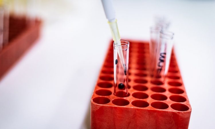
By definition, flow cytometry refers to the immunophenotyping technique in which cell suspensions are stained with specific, fluorescently labeled antibodies. These cell suspensions can be derived from a wide range of sample types, including blood, bone marrow, cerebrospinal fluid (CSF), pleural fluid, microorganisms, soluble antigens, and even solid tissues. Once injected into the flow cytometer, this technique can provide a great deal of information on the cells in the sample.
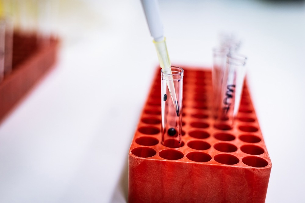
Image Credit: mh_enders/Shutterstock
For example, flow cytometry can distinguish different cellular properties from morphology to cell cycle stage. Furthermore, the information provided by a flow cytometer reflects the individual cells within the population rather than providing an average that is often the result of other molecular biology techniques like Western blotting.
Flow cytometry and disease physiology
The first clinical application of flow cytometry was reported in the late 1980s when it was used to manage patients with the human immunodeficiency virus (HIV). Since then, flow cytometry has become an indispensable tool for various medical fields, particularly in hematology, immunology, and clinical pathology.
Immunological diseases
Primary immunodeficiencies (PID) can be classified into nine categories, which include severe and combined immunodeficiencies (SCID), combined immunodeficiencies with syndromic features, predominantly antibody deficiencies, immunodysregulated disorders, defects affecting phagocytes, defects of innate immunity, autoinflammatory disorders, complement deficiencies, and phenocopies of PIDs. Typically, severe PIDs will cause symptoms to develop shortly after birth; however, the precise diagnosis of these disorders requires specific and sensitive tests.
As a result, flow cytometry is often considered the method of choice for both diagnosing and studying PIDs. Typically, a flow cytometry assay is employed after a clinician assesses the clinical presentation of the patient and runs certain basic laboratory tests. If the healthcare provider suspects a defect of adaptive immunity, a basic lymphocyte phenotyping will then be performed to assess the number and proportion of immune cells, as well as provide an overview of the cellular processes of the patient, including cellular proliferation, cytokine secretion, and cytotoxicity, to name a few.
Flow cytometry can also be used to assess the subpopulations of different immune cells to understand better the physiology of a given patient’s disease state. For example, if a clinician is interested in assessing a patient’s lymphocyte subpopulations, they can request a flow cytometry assay on the CD4+ and CD8+ T-cells, as well as information on the B-cells and natural killer (NK) cells within the patient’s sample. Aside from its utility in diagnosing a primary or secondary immunodeficiency, flow cytometry used to assess lymphocyte subpopulations can also be particularly useful in monitoring how patients respond to certain immunotherapies, such as the anti-CD20 antibody treatment known as rituximab.
Comparatively, extended phenotypic analysis can better understand the behavior of dendritic cell subpopulations, regulatory T-cells, and recent thymic emigrants.
Hematology and oncology
One of the most prominent applications of flow cytometry for disease physiology purposes can be found in hematology and oncology, particularly when a hematopoietic neoplasm is suspected in blood, bone marrow, or tissue biopsy samples. To this end, a neoplastic cell is identified through flow cytometry by quantitatively measuring changes that arise in cell subset distribution of qualitative antigen expression changes.
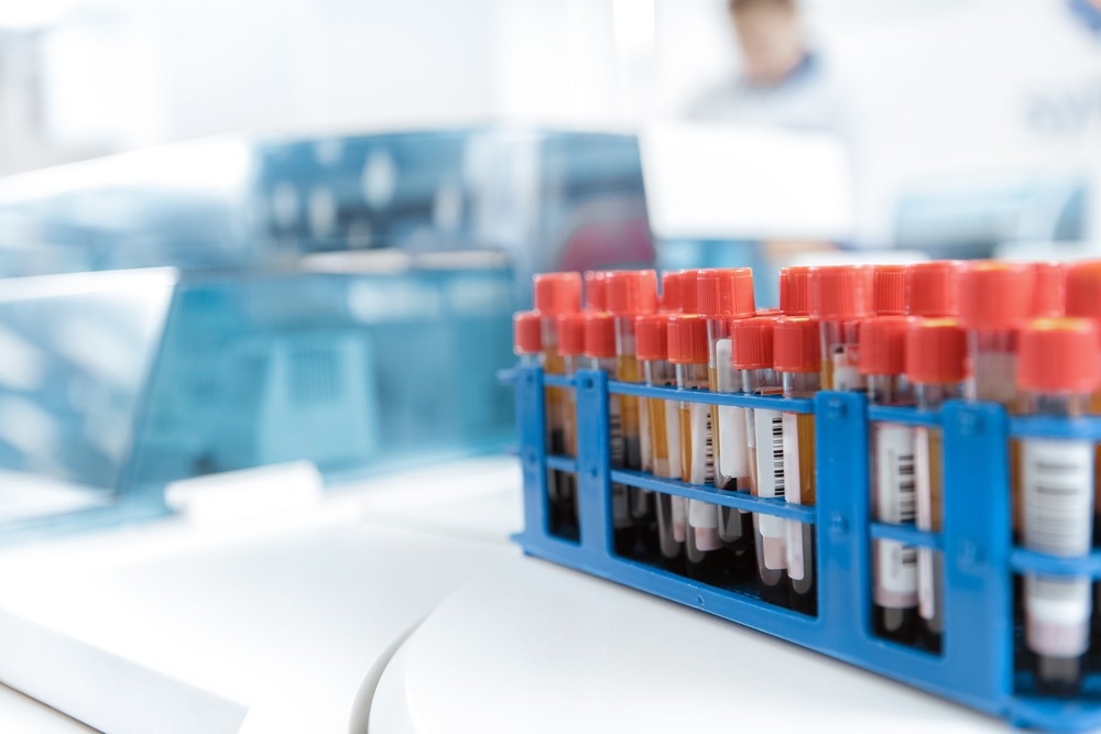
Image Credit: Nestor Rizhniak/Shutterstock
For example, in the case of B cell lymphoma, flow cytometry is useful in determining the clonality of B cells, which often equates with neoplasia. Neoplastic T-cells that indicate T-cell lymphomas and lymphoproliferative processes can also be assessed by flow cytometry of T-cell subsets and/or an assessment of T-cell antigen expression.
Samples derived from patients suspected of having a platelet disorder are also often subject to flow cytometry. For example, flow cytometry is often used to determine the presence of autoantibodies to platelets that are often indicative of immune thrombocytopenia. This approach is considered superior to other laboratory tests that detect antiplatelet antibodies, particularly those that are low-density or labile. Several other platelet disorders, including Glanzman’s thrombasthenia and Bernard-Soulier syndrome, can also be diagnosed and monitored through flow cytometry by assessing the presence of antigens on the surface of these platelets.
Conclusion
Although flow cytometry is often considered to be primarily a diagnostic tool, it is also widely used to monitor the efficacy of treatments and the progression of certain diseases. Together, these flow cytometry applications contribute to a better global understanding of various health conditions to ultimately improve patient outcomes for the future.
References
- Virgo, P. F., & Gibbs, G. J. (2011). Flow cytometry in clinical pathology. Annals of Clinical Biochemistry: International Journal of Laboratory Medicine. doi:10.1258/acb.2011.011128.
- Salzer, U., Sack, U., & Fuchs, I. (2019). Flow Cytometry in the Diagnosis and Follow Up of Human Priamry Immunodeficiencies. EJIFCC 30(4); 407-422. https://www.ncbi.nlm.nih.gov/pmc/articles/PMC6893889/.
- Meyerson, H. (2018). Flow Cytometry in Hematology. Concise Guide to Hematology 253-275. doi:10.1007/978-3-319-97873-4_22.
Further Reading
- All Flow Cytometry Content
- Using Flow Cytometry in Disease Diagnosis
- Flow Cytometry and Drug Discovery
- Using Flow Cytometry in Biomarker Detection
- Flow Cytometry Methodology, Uses, and Data Analysis
Last Updated: Sep 26, 2022

Written by
Benedette Cuffari
After completing her Bachelor of Science in Toxicology with two minors in Spanish and Chemistry in 2016, Benedette continued her studies to complete her Master of Science in Toxicology in May of 2018.During graduate school, Benedette investigated the dermatotoxicity of mechlorethamine and bendamustine; two nitrogen mustard alkylating agents that are used in anticancer therapy.
Source: Read Full Article