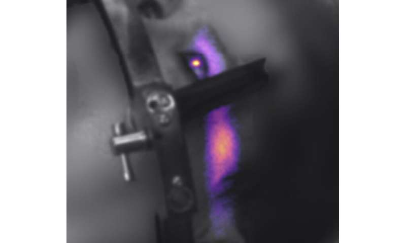
For decades, theories have existed to explain patient reports of seeing light flashes during radiation therapy, even with their eyes closed. However, no one has been able to secure evidence of this sensation in humans to prove their theory. A team of researchers at Dartmouth’s and Dartmouth-Hitchcock’s Norris Cotton Cancer Center led by radiation oncologist Lesley Jarvis, MD, Ph.D. and Irwin Tendler, MEng, have collected real-time data to show that during head and neck radiation therapy, enough light is produced inside the eye to elicit such a visual sensation. This evidence, and their explanation of it, “Experimentally Observed Cherenkov Light Generation in the Eye During Radiation Therapy,” is newly published in The Red Journal (International Journal of Radiation Oncology, Biology, & Physics).
“Overall, the idea of imaging light emission from humans undergoing radiotherapy is novel and was also first suggested and developed at Dartmouth,” says Tendler. “Our newest data is exciting because for the first time, light emission from the eye of a patient undergoing radiotherapy was captured. This data is also the first instance of evidence directly supporting that there is enough light produced inside the eye to cause a visual sensation and that this light resembles Cherenkov emission.”
Light emission from the eye is very subtle, hard to detect, and likely why no one has been able to record evidence of this phenomenon. To accomplish this, the team leveraged special technology called the CDose camera imaging system, an engineering innovation manufactured by Dartmouth spin-off company, DoseOptics, LLC. The camera provides live views of light emission from biological systems (animal and human) during radiotherapy. “As the radiation beam passes through the eye, light is generated within the vitreous fluid. Our real-time data rigorously showed that the amount of light produced is sufficient to elicit a visual sensation—a topic that has been debated in the literature,” explains Tendler. “By analyzing the spectral composition, we also show that this emission can be classified as Cherenkov light—again, another contested point in the literature.”
The benefits of the team’s finding to cancer treatment are several-fold:
- The imaging technique could be used to monitor light emission from the eye during radiotherapy and directly determine whether radiation transects the eye. “In the case that the eye is a target, the method could provide confirmation of beam delivery; in the case that this is unintentional, it can provide evidence of an error or near miss—as a safety check,” says Tendler.
- The ability to provide the patient with a valid explanation of this phenomenon can help relieve treatment-related anxiety.
- “Published work has shown that if a patient doesn’t see light flashes during radiotherapy, there is a higher chance of expecting vision loss after irradiation,” says Tendler. The method could help determine if any light was actually generated to potentially relate this to predicted vision loss as well as provide information about long-term visual outcome following radiotherapy.
Source: Read Full Article
