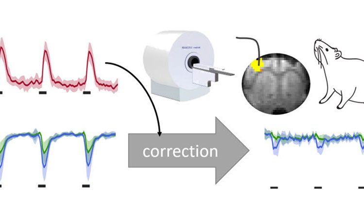
 sensors is feasible. For detection of lactate levels, correction of hemodynamic artifacts is required, which can be achieved by using simultaneously or separately acquired functional magnetic resonance imaging (fMRI) signals. Credit: H. Lambers, University of Münster.")
Lactate is generated in the body as a byproduct during normal metabolism and exercise. While considered a useless waste product for a long time, recent studies have shown that lactate can be an indicator for cancer and various other diseases. In light of this, monitoring lactate levels can help us gain a better understanding of the energy metabolism process. However, current detection methods provide recordings of bulk lactate levels outside the cell only and not enough temporal resolution to detect real-time changes in these levels. This makes it particularly challenging to determine lactate levels in the brain where their concentrations are lower than other metabolites.
To improve the detection sensitivity of lactate, a lactate sensor with high temporal resolution known as “Laconic” has recently been developed. The sensor contains two fluorescent chemical compounds (or “fluorophores”), termed “donor” and “acceptor,” and is based on a mechanism called “fluorescence resonance energy transfer” (FRET). These compounds emit light of different wavelengths and are separated by a molecule that is capable of interacting with lactate. When the sensor is irradiated with light, the donor absorbs the energy and transfers it to the acceptor molecule. The intensity of the emitted light depends on the energy transfer and the distance between the two fluorophores. When lactate interacts with the molecule separating the fluorophores, it changes the distance between them and, in turn, the intensity of the light emitted. Therefore, these sensors can detect the presence of lactate by observing the signal intensity of the emitted light. Together with a functional MRI scan, this can help map cell-specific lactate levels and link them to the blood flow in the brain. However, changes in blood flow can introduce artifacts in the measured signals. To detect minute lactate levels, it is necessary to identify and correct for these artifacts.
On this front, researchers from University Hospital Münster, Germany, and Paris-Saclay University, France, recently investigated how these sensors respond to the conditions in the brain, specifically to the changes in blood flow and blood oxygen levels. The results of the study are published in the journal Neurophotonics.
Along with Laconic, the researchers used a FRET-based calcium sensor for examining the artifacts. The Laconic sensors were genetically encoded in rat models to be expressed in the sensory cortex, the part of the brain that receives sensory information. To obtain fluorescence signals, an optical fiber was implanted just above the sensor to guide the light going in and out of the brain region under observation. Lactate and calcium concentrations in the brain were then measured following the stimulation of the sensory cortex and intravenous (IV) injections of lactate.
Both the calcium and lactate sensors responded to the changes in brain activity. The lactate sensors expressed in the neurons (brain cells) were able to detect increases in lactate concentration introduced by the IV injections. However, the recordings were affected by the hemoglobin in the blood, which also absorbed light along with the fluorophores. Since changes in blood flow modified the hemoglobin concentration, it caused a change in the intensity of fluorescence signals as well. The researchers corrected for these artifacts by using a correction algorithm that removed them for both simultaneous and separate measurements of fluorescence and MRI signals. Additionally, they validated their measurements against local field potential recordings and blood analyses.
Source: Read Full Article