
Scientists believe that an unbiased analysis of antibody binding sites would provide significant insights into health and disease states. In this regard, many researchers have utilized programmable phage display libraries to detect novel autoantibodies. Phage display libraries have been used to characterize anti-viral immunity and profile allergen-specific Immunoglobulin E (IgE) antibodies.
Even though protein microarrays are extremely useful for protein research, the high per-assay cost and numerous technical artifacts make them less user-friendly. Technologies such as Nucleic Acid Programmable Protein Array (NAPPA) and single-molecule PCR-linked in vitro expression (SIMPLEX) are effective but not cost-effective.
To overcome the limitations of array-based profiling of full-length proteins, scientists developed a novel methodology known as Parallel Analysis of Translated Open reading frames (PLATO), based on ribosome display of open reading frame (ORF) libraries. However, some limitations of PLATO have hindered its mass acceptance in the scientific community.
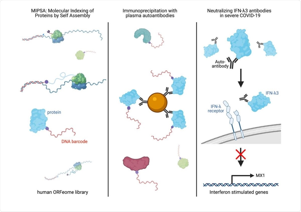
Now, scientists have recently developed a novel alternative molecular display technology called Molecular Indexing of Proteins by Self Assembly (MIPSA). Their research, published on the bioRxiv* preprint server, explains how this technology overcomes the critical disadvantages of PLATO and other full-length protein array technologies.
MIPSA produces libraries of soluble full-length proteins. Each protein can be identified explicitly by covalent conjugation to a DNA barcode lined by universal PCR primer binding sequences. Researchers have incorporated the barcodes at the 5’ end of transcribed messenger RNA (mRNA) sequences that is positioned upstream of the ribosome binding site (RBS). A cDNA barcode is produced by reverse transcription (RT) of the 5’ end of in vitro transcribed RNA (IVT-RNA) connected to a haloalkane-labeled RT primer. An N-terminal HaloTag is merged to the protein encoded downstream of the RBS. This ensures in vitro translation in the intra-complex (“cis”) covalent coupling of the cDNA barcode to the HaloTag and its downstream open reading frame (ORF) encoded protein product. This newly formed library containing uniquely indexed full-length proteins can be explored in many proteome studies, e.g., unbiased autoantibody profiling. The current research has unveiled this platform's utility by revealing the presence of known and novel autoantibodies in the plasma of severely affected COVID-19 patients.
A MIPSA Gateway Destination vector has been developed using several vital ingredients. It contains a T7 RNA polymerase transcriptional start site, an isothermal unique clonal identifier (“UCI”) barcode sequence flanked by constant primer binding sites, a ribosome binding site (RBS), an N-terminal HaloTag fusion protein, recombination sequences for ORF insertion, a stop codon, and a homing endonuclease site for plasmid linearization.
The main advantage of MIPSA over various other technologies such as protein microarrays, SIMPLEX, NAPPA and PLATO is that this system provides a novel molecular display for full-length proteins. Further, MIPSA offers a simple, high throughput, and cost-effective sequencing library that provides stability to the protein-DNA complexes. These attributes are incredibly beneficial for both the manipulation and storage of display libraries.
MIPSA facilitates unbiased analyses of protein-antibody, protein-protein, and protein-small molecule interactions. It also contributes to post-translational modification studies, for example, hapten modification studies and protease activity profiling. Further, this technology can be easily adopted by standard molecular biology laboratories. This is because it does not require sophisticated equipment or training. Availability of a simple high throughput DNA sequencing instrument is the only essential requirement.
Even though the MIPSA display system utilizes HaloTag/HaloLigand system, it can work with the SNAP-tag forming a covalent bond with benzylguanine (BG) derivatives. BG could be utilized as a label to RT primer replacing HaloLigand. MIPSA can also support mutant derivative of the SNAP-tag, and the CLIP-tag that binds O2-benzylcytosine derivatives.
Scientists have reported the association of autoimmunity with severe COVID-19 disease. The current research involves the detection of multiple autoantibodies using the MIPSA display system. In patients with severe COVID-19 disease, neutralizing IFN-α/ω autoantibodies have been reported. These autoantibodies infrequently occur in the general population and block the immune response to viral replication in cells.
MIPSA complements techniques like PhIP-Seq. One limitation of this technology is that the MIPSA protocol requires cap-independent and cell-free translation. It is expected that future work will help in overcoming this drawback. In the current study, scientists have used MIPSA to detect some known autoantibodies and discover neutralizing Interferon lambda 3 (IFN-λ3) autoantibodies.
In conclusion, the researchers suggest that IFN-λ3 autoreactivity may be more frequent among individuals with severe COVID-19. This is the first report describing neutralizing IFN-λ3 autoantibodies and therefore proposes a potentially novel pathogenic mechanism contributing to life-threatening COVID-19 in a subset of patients. This discovery would help identify individuals who are at a higher risk of SARS-CoV-2 infection and, thereby, promote the therapeutic usage of interferon-beta in this vulnerable group.
*Important Notice
bioRxiv publishes preliminary scientific reports that are not peer-reviewed and, therefore, should not be regarded as conclusive, guide clinical practice/health-related behavior, or treated as established information.
- Neutralizing IFNL3 Autoantibodies in Severe COVID-19 Identified Using Molecular Indexing of Proteins by Self-Assembly, Joel J. Credle, Jonathan Gunn, Puwanat Sangkhapreecha, Daniel R. Monaco, Xuwen Alice Zheng, Hung-Ji Tsai, Azaan Wilbon, William R. Morgenlander, Yi Dong, Sahana Jayaraman, Lorenzo Tosi, Biju Parekkadan, Alan N. Baer, Mario Roederer, Evan M. Bloch, Aaron A. R. Tobian, Israel Zyskind, Jonathan I. Silverberg, Avi Z. Rosenberg, Andrea L. Cox, Tom Lloyd, Andrew L. Mammen, H. Benjamin Larman, bioRxiv, 2021.03.02.432977; doi: https://doi.org/10.1101/2021.03.02.432977, https://www.biorxiv.org/content/10.1101/2021.03.02.432977v1
Posted in: Medical Science News | Medical Research News | Miscellaneous News | Disease/Infection News | Healthcare News
Tags: Allergen, Antibodies, Antibody, Assay, Autoantibodies, Autoimmunity, Cell, Codon, Conjugation, Coronavirus Disease COVID-19, DNA, DNA Sequencing, Endonuclease, High Throughput, Immune Response, Immunoglobulin, in vitro, Molecular Biology, Molecule, Nucleic Acid, Plasmid, Polymerase, Protein, Proteome, Research, Ribosome, RNA, SARS, SARS-CoV-2, Transcription, Translation

Written by
Dr. Priyom Bose
Priyom holds a Ph.D. in Plant Biology and Biotechnology from the University of Madras, India. She is an active researcher and an experienced science writer. Priyom has also co-authored several original research articles that have been published in reputed peer-reviewed journals. She is also an avid reader and an amateur photographer.
Source: Read Full Article