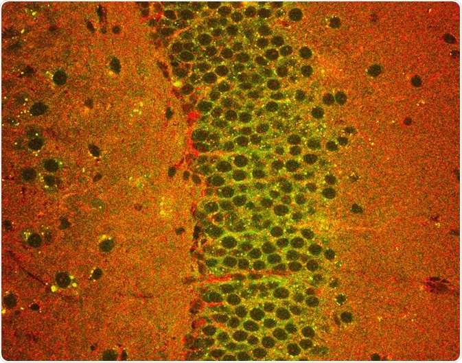
Mouse brain slices are widely used in neurological research. Scientists have long been interested in using these samples to understand proteome changes in response to neuroactive compounds. However, the protein extraction yield from brain slices is usually too low for protein analysis.
In this case study, researchers demonstrated a two-phase extraction method that allows brain slice protein analysis by nano liquid chromatography coupled to ion mobility mass spectrometry (nanoLC-MS), with particular emphasis on membrane proteins.

Much of what we know about the function of the brain has come from neurological studies on mouse brain slices, which are usually thinner than 0.5 mm. Such slices maintain the natural environment for the brain cells inside but are easier to access experimentally than the full brain.
They can be kept functional for weeks in a physiological nutrient solution to carry out neurological experiments, such as measurements of the electrical activity of the brain slice neurons, tests of neurotoxins and neuroactive drugs, or experiments observing cellular signaling in brain tissues.
In all these cases, membrane proteins, such as ion channels or receptor proteins, play an important role, but these proteins are difficult to analyze as protein extractions from the thin slices are usually low in yield.
In previous research, the brain slice proteins had been purified and enriched by methods such as the biotin-streptavidin pull-down system. In this method, proteins are tagged with biotin by chemical or enzymatic reaction at specific protein sites.
In parallel, streptavidin is linked to a solid support. When the biotin-tagged protein fraction passes through that support, the linked streptavidin binds the biotin and thus pulls the tagged protein out of solution.
While this method is well established, it does not discriminate water-soluble and membrane proteins. This discrimination can be achieved by additional methods such as density gradient ultracentrifugation, which however requires long centrifugation times.
A recent study from investigators at the University Medical Center Rostock (Germany), led by Professor Oliver Schmitt, presents an alternative membrane protein enrichment method based on an aqueous two-phase (biphasic) polymer system and demonstrates its use for mouse brain slice proteomics.
Sorting Protein Fractions
The researchers used a mixture of dextran (a sugar polymer) and PEG (polyethylene glycol) to separate proteins from a homogenized brain slice suspension. Adding the two polymers together in an aqueous solution creates a hydrophilic dextran phase at the bottom, while a rather hydrophobic PEG phase forms at the top.
In effect, hydrophobic membrane fractions from the brain slice suspension primarily locate in the PEG phase, while water-soluble proteins preferentially reside in the dextran phase. Membrane fractions were recovered from the PEG phase by ultracentrifugation, and dissolved in urea plus detergent solution.
To prepare for proteomics analysis by LC-MS, the samples were desalted with ultrafiltration columns which retain the protein fraction while allowing salts to pass through. Proteins were then digested into peptide fragments by on-column protease digestion with trypsin.
Subsequent proteomics analyses were carried out on a nano liquid chromatography (nanoLC) system coupled to a high-definition tandem mass spectrometer (HDMS) which was equipped with an ion mobility separator for enhanced mass spectrometry resolution.
Insights into the Brain Slice Proteome
The researchers identified 1,161 proteins, of which 369 (32 %) were membrane proteins and 792 (68 %) were not membrane-associated or could not be classified. This demonstrated that the extraction method was suitable for LC-MS proteomics of the small brain slice samples.
The presence of non-membrane proteins is not unexpected since the two-phase extraction method does not fully discriminate the protein classes. However, only a relative enrichment is required to improve the signals of the membrane protein fraction by lowering the signals from soluble proteins.
This method can now be employed for proteomics studies of brain slices after applying various stimuli, for instance, a neurotoxin. However, it remains a challenge to compare the proteomics profile of stimulated slices against non-stimulated controls, as it is difficult to keep individual slices under exactly the same conditions except for one stimulus.
In this study, the authors made a step forward by comparing brain slices of different animals from the same inbred mouse line, and they reported a reasonably low variance of abundance for most proteins. This suggests that stimulated samples and non-stimulated controls could be prepared as distinct brain slices, though further studies will need to validate these findings.
Sources
- Joost S et al. (2019). Membrane Protein Identification in Rodent Brain Tissue Samples and Acute Brain Slices. Cells. DOI: 10.3390/cells8050423
- Humpel C. (2015). Organotypic brain slice cultures: A review. Neuroscience. DOI: 10.1016/j.neuroscience.2015.07.086.
Further Reading
- All Neuroscience Content
- What is Neurology?
- What is the Difference between Neurology and Neuroscience?
- What is Neuroscience?
- What is Neurosurgery?
Last Updated: Sep 9, 2019

Written by
Christian Zerfaß, Ph.D.
Christian is an enthusiastic life scientist who wants to understand the world around us. He was awarded a Ph.D. in Protein Biochemistry from Johannes Gutenberg University in Mainz, Germany, in 2015, after which he moved to Warwick University in the UK to become a post-doctoral researcher in Synthetic Biology.
Source: Read Full Article
