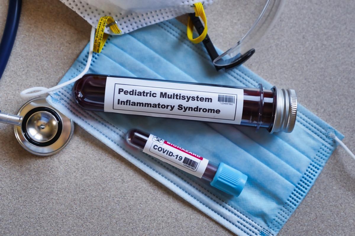
In a recent study published in Biology* researchers determined the immune signature of the multisystem inflammatory syndrome in children (MIS‐C) among hospitalized children diagnosed with severe acute respiratory syndrome coronavirus 2 (SARS‐CoV‐2) infection.

MIS-C is a rare and delayed complication that develops after three to eight weeks of SARS‐CoV‐2 infection. It is characterized by a hyperinflammatory state and multiorgan dysregulation. Although the clinical presentation of MIS-C is similar to other inflammatory syndromes, such as cytokine storm syndrome (CSS), primary hemophagocytic lymphohistiocytosis (pHLH), and sepsis, it remains unclear why only a few COVID-19 affected children are predisposed to MIS-C.
About the study
In the present retrospective study, the researchers screened MIS-C patients for genomic variations in several immunoregulatory genes to determine the genetic association of MIS-C with SARS‐CoV‐2.
A total of 45 children suspected of MIS-C were hospitalized at the Northwell Cohen Children’s Medical Center between April 17 and July 9, 2020. Of these, 39 children meeting the Centers for Disease Control and Prevention (CDC) criteria of MIS-C were allocated as Group 1 whereas the remaining children comprised Group 2 or the control group.
Blood samples were obtained from all the participants in the first follow-up outpatient visit post-hospital discharge. These samples were subjected to genetic screening using a monogenic autoimmune panel comprising 109 genes of immunological dysfunction. The genetic analysis was performed based on the American College of Medical Genetics and Genomics guidelines. Additionally, public databases were used to calculate the population frequencies.
Genetic variations in the dedicator of cytokinesis 8 (DOCK8) gene were assessed in vitro for all participants since this gene was most prevalent in the study cohort. Peripheral blood mononuclear cells (PBMC) containing DOCK8 messenger ribonucleic acid (mRNA) were obtained from healthy donors to generate complementary deoxyribonucleic acid (cDNA) by reverse transcription-polymerase chain reaction (RT-PCR). Patient cDNA copies equivalent to DOCK8 mutant sequences were produced by site-directed mutagenesis and were validated using the Sanger DNA sequence analysis.
Additionally, human natural killer 92 (NK92) cells were transduced with a recombinant human foamy virus (FV) to evaluate the effect of DOCK8 mutation on NK cell function. The NK cells were analyzed for cell death using anti-CD56 (cluster of differentiation 56), an NK cell marker, by flow cytometry. The K562 erythroleukemia target cells were used as controls.
Results and discussion
Based on the genetic analysis, Group 1 children were further classified as Group 1a comprising children with the absence of variants of uncertain significance (VUS) (n=10), Group 1b comprising children with non-pHLH/DOCK8 VUS (n=19), and Group 1c comprising children with VUS either in DOCK 8 or in other pHLH genes (n=10). Moreover, a subset of Group 1c.1 comprised children with DOCK8 VUS (n=4).
The average age of MIS-C children was nine years and most of them (62%) were males. They were hospitalized for an average of seven days. About 64% of the MIS-C children required pediatric intensive care unit (PICU) admissions while 49% of the children needed oxygen supplementation. Notably, none of the controls has these requirements. Additionally, thrombocyte counts and ferritin were elevated in Group 1b children. However, these serological findings were not statistically significant.
The results showed that 54 rare VUS (prevalence <0.1%) were detected among the immunological genes in Group 1. About 74% of the MIS-C children had a minimum of one variant. Among these genomic variations, missense mutations (amino acid alterations) in the DOCK8 gene were the most commonly observed mutations. Additionally, 15% of children demonstrated heterozygous and rare missense mutations in pHLH genes (LYST, STXBP2, PRF1, AP381, and UNC13D). One patient had missense mutations in the NLR family CARD domain containing 4 (NLRC4) genes, responsible for CSS autoimmunity. Notably, genetic variations in the pHLH and DOCK8 genes were detected in the MIS-C group only (25%).
The four missense mutations in the DOCK8 genes decreased the NK cell degranulation and cytotoxicity compared to wild-type (WT) DOCK8 in vitro. Additionally, the NK cells expressing DOCK8 mutations had a substantially lower CD107a expression than WT DOCK8-transduced NK cells. Likewise, reduced cytotoxicity against the K563 target cells was noted among the mutant DOCK8-expressing NK cells.
To conclude, genetic screening enables the detection of mutant immunoregulatory genes causing MIS-C among COVID-19-infected children, which could identify children at high risk and facilitate prompt treatment. However, further research is needed for better elucidation of MIS-C pathophysiology.
- Vagrecha, A.; Zhang, M.; Acharya, S.; Lozinsky, S.; Singer, A.; Levine, C.; Al‐Ghafry, M.; Fein Levy, C.; Cron, R.Q. (2022). Hemophagocytic Lymphohistiocytosis Gene Variants in Multi‐System Inflammatory Syndrome in Children. doi: https://doi.org/10.3390/ biology11030417 https://www.mdpi.com/2079-7737/11/3/417
Posted in: Medical Science News | Medical Research News | Disease/Infection News | Healthcare News
Tags: Amino Acid, Autoimmunity, Blood, Cell, Cell Death, Children, Coronavirus, Coronavirus Disease COVID-19, covid-19, Cytokine, Cytometry, Cytotoxicity, DNA, Flow Cytometry, Gene, Genes, Genetic, Genetics, Genomic, Genomics, Hemophagocytic Lymphohistiocytosis, Hospital, in vitro, Intensive Care, Mutation, Oxygen, Pathophysiology, Polymerase, Polymerase Chain Reaction, Research, Respiratory, Ribonucleic Acid, SARS, Sepsis, Severe Acute Respiratory, Severe Acute Respiratory Syndrome, Syndrome, Transcription, Virus

Written by
Pooja Toshniwal Paharia
Dr. based clinical-radiological diagnosis and management of oral lesions and conditions and associated maxillofacial disorders.
Source: Read Full Article
