In a recent study published in the Immunity journal, researchers explored the current knowledge of the mechanism employed by messenger ribonucleic acid (mRNA) vaccines to elicit innate immune activation.
The nucleoside-modified mRNA vaccines used against coronavirus disease 2019 (COVID-19) include BNT162b2 (Pfizer/BioNTech) and mRNA-1273 (Moderna). These nucleoside-modified mRNA- lipid nanoparticles containing ionizable lipids (iLNPs) vaccines have shown high effectiveness at generating spike-specific adaptive immune responses, especially neutralizing antibodies for the induction of protective immunity against COVID-19. However, further research is essential to understand the various components of innate immunity required for generating protective immune responses.
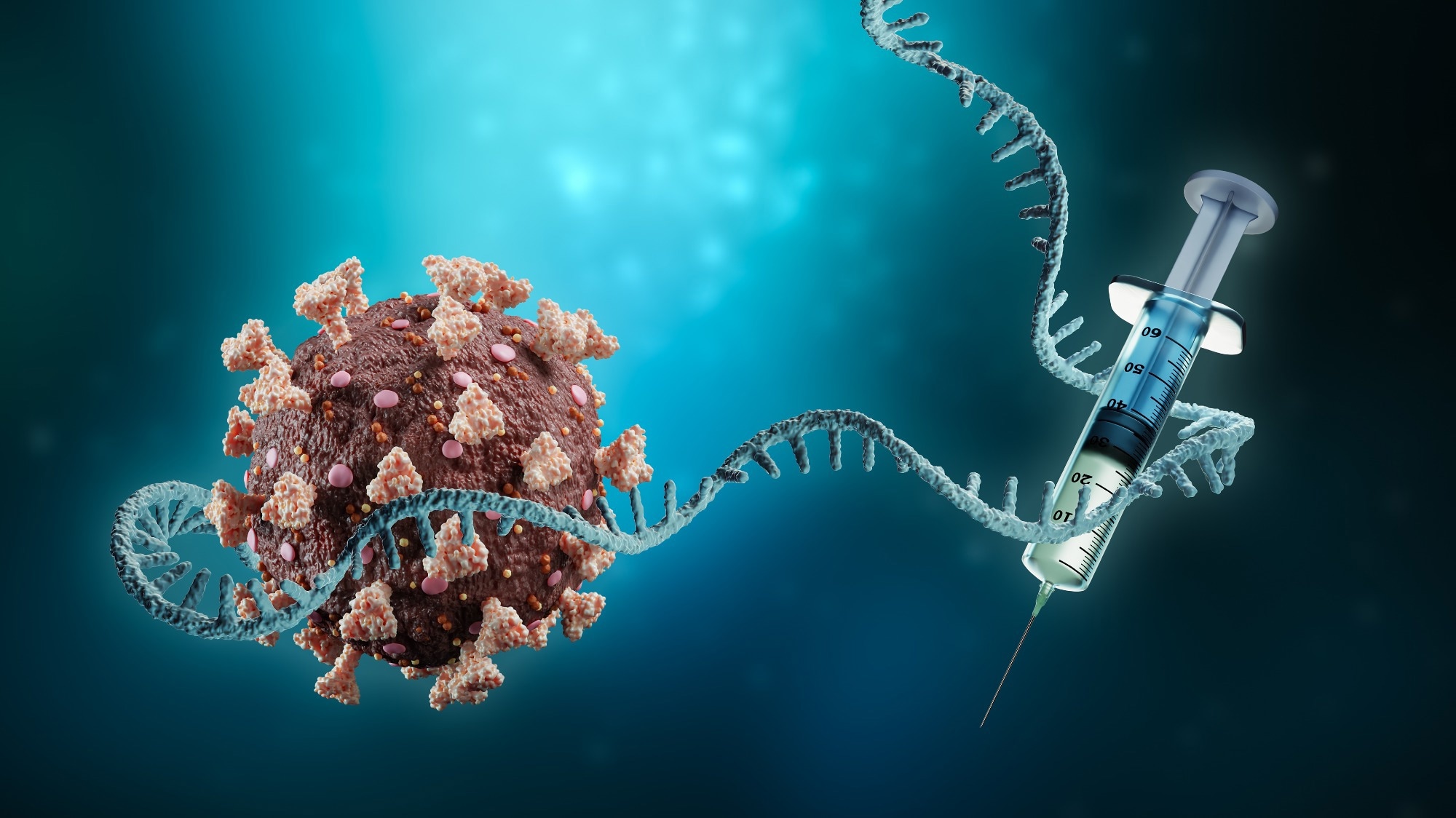
 Study: Innate immune mechanisms of mRNA vaccines. Image Credit: MattLphotography / Shutterstock
Pharmacokinetic profile of nucleoside-modified mRNA-iLNP vaccines
mRNA-iLNP vaccines disperse in the body based on the formulation and the mode of delivery of iLNP. COVID-19 mRNA vaccine administration in the human deltoid muscle likely resulted in antigen production observed at the injection site and draining apical, axillary, and supraclavicular lymph nodes (LNs). Comparable biodistribution is observed for standard vaccination types, like adjuvanted protein subunits.
Depending on the mRNA dose, the encoded protein, the iLNP type, and the method of mRNA-iLNP delivery, protein synthesis after mRNA-iLNP injection begins early and peaks rapidly after four to 24 hours before declining gradually. These events may extend for a couple of days or over a week. Spike-encoding mRNA and spike protein are detected in germinal centers (GCs) present in the draining axillary LNs of vaccinated people for up to 60 days following the second mRNA-1273 or BNT162b2 vaccine dose. However, the time span for which antigen expression lasts requires more investigation.
Nucleoside-modified mRNA-iLNP vaccines generate significant T follicular helper (Tfh) cell responses while forming GCs required to produce antibodies with high persistence and affinity. In the draining LNs of patients vaccinated with BNT162b2, Tfh and GC-B cell responses remain up to six months after the second mRNA dose before culminating in the production of affinity-matured memory B cells with bone marrow-resident plasma cells. COVID-19 mRNA vaccines generate antigen-specific Tfh cells, CD4+ T cells, T helper 1 (Th1) polarization, and IFN-g-producing CD8+ T cells that are traceable for up to six months following booster vaccination.
The majority of immune cells that infiltrate injection sites are neutrophils. Neutrophils can easily internalize fluorescently labeled iLNPs, yet their synthesis of the encoded reporter protein is inadequate. Therefore, it is possible for neutrophils to contend with other immune cells for mRNA-iLNP vaccine uptake. On the contrary, dendritic cells (DCs) subsets and monocytes effectively acquire and translate mRNA-iLNPs. Parallelly, the team observed that single-cell RNA-sequencing examination of draining LNs from BNT162b2-vaccinated mice detected the presence of spike mRNA within myeloid DC, macrophage, and monocyte cells.
Lab Diagnostics & Automation eBook

Activated monocytes, DCs, and macrophages are among the essential cell types that cause mRNA-iLNP absorption, protein synthesis, and presentation of antigens in the lymphoid tissue to facilitate the adaptive immune response.
Immunosensitivity to unmodified and nucleoside-modified mRNA
The three basic types of innate immune sensing for synthetic mRNA include (1) uridine-dependent identification of different RNA species, (2) detection of double-stranded RNA (dsRNA), and (3) identification of the 5' end of mRNA if it is not correctly capped. Unfortunately, there is currently a significant lack of research directly assessing the immunogenicity and effectiveness of different mRNA vaccination platforms.
Modified nucleosides can inhibit the activation of numerous dsRNA sensors, such as Toll-like receptor 3 (TLR3), 2'-5'-oligoadenylate synthetase (OAS), protein kinase R (PKR), melanoma differentiation-associated protein 5 (MDA5), and retinoic acid-inducible gene I (RIG-I). In addition, the detection of uridine-containing RNA is correlated with enhanced expression of pro-inflammatory cytokines, specifically type-I interferons (IFN), which stimulate RNA sensor expression further and can block the expression of antigen from the mRNA through the actions of PKR and OAS.
Even though modified nucleosides have certain benefits, unmodified mRNA is equally capable of achieving significant protein expression and antibody neutralization responses. CureVac devised a sequence-engineering technique to evade detection via pattern recognition receptors by modifying the codon use with synonymous substitutions that limit uridine and enhance GC content, encouraging robust antigen expression and immunogenicity.
Next-generation mRNA-iLNP vaccines
Considering the mRNA component, the m1J nucleoside alteration appears to enhance mRNA-iLNP vaccine tolerance in humans by reducing innate immune recognition, with potential advantages related to adaptive immunity and translation efficiency. Furthermore, several investigations demonstrate that nucleoside-modified mRNA-iLNP vaccines can effectively produce a temporary type I IFN response, indicating the potential for complementary or synergistic activation of the innate immune system with the mRNA component. Interestingly, this showed that the MDA5-IFN-a signaling pathway is essential for the CD8+ T cell response to vaccination of mice with BNT162b2.
Overall, the study findings showed that during the COVID-19 pandemic, the nucleoside-modified mRNA-iLNP platform represented a significant breakthrough in vaccine development that proved to be a boon in the middle of the pandemic. The researchers anticipate that these advancements will result in the evolution of mRNA-iLNPs having increased efficacy, tolerability, and safety compared to mRNA-1273 and BNT162b2.
- Innate immune mechanisms of mRNA vaccines, Rein Verbeke, Michael J. Hogan, Karin Loré, Norbert Pardi, Immunity, VOLUME 55, ISSUE 11, DOI: https://doi.org/10.1016/j.immuni.2022.10.014, https://www.cell.com/immunity/fulltext/S1074-7613(22)00555-6
Posted in: Medical Science News | Medical Research News | Disease/Infection News
Tags: Antibodies, Antibody, Antigen, B Cell, Bone, Bone Marrow, CD4, Cell, Codon, Coronavirus, Coronavirus Disease COVID-19, Cytokines, Efficacy, Evolution, Gene, Immune Response, Immune System, immunity, Interferons, Kinase, Lipids, Lymph Nodes, Macrophage, Melanoma, Monocyte, Muscle, Nanoparticles, Neutrophils, Nucleoside, Pandemic, Protein, Protein Expression, Protein Synthesis, Receptor, Research, Retinoic Acid, Ribonucleic Acid, RNA, Signaling Pathway, Spike Protein, Translation, Vaccine

Written by
Bhavana Kunkalikar
Bhavana Kunkalikar is a medical writer based in Goa, India. Her academic background is in Pharmaceutical sciences and she holds a Bachelor's degree in Pharmacy. Her educational background allowed her to foster an interest in anatomical and physiological sciences. Her college project work based on ‘The manifestations and causes of sickle cell anemia’ formed the stepping stone to a life-long fascination with human pathophysiology.
Source: Read Full Article
