New imaging of patients with Alzheimer’s demonstrates how a telltale protein spreads throughout the brain based on the phenotype of the disease, i.e., whether the condition is dominated by forgetfulness, or atrophy in a specific brain region. The research offers a host of illuminating clues that ultimately may inform new treatment strategies.
The protein is known as tau and a large multi-disciplinary team of brain researchers at McGill University in Montreal has been able to trace the protein’s patterns in living patients via magnetic resonance imaging (MRI). Alzheimer’s disease is intimately linked to tau, which can form tangles in the brain that irrevocably damage neurons.
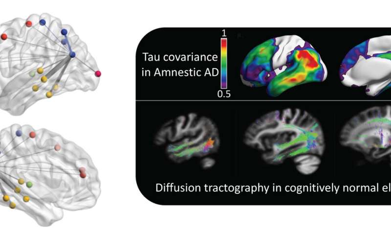
The patterns detected by McGill scientists apparently are unique to the phenotype of Alzheimer’s afflicting the patient. This staggering finding opens an intriguing new window into the molecular mechanisms of the disease. And while many features of Alzheimer’s are the same from one patient to the next, phenotypes are a hallmark of the condition. Tracking tau patterns is a specialty of the scientists at McGill, who found that the intrinsic connectivity of the human brain itself provides the scaffolding for the aggregation of tau in distinct variants of the disease.
“Although some of the symptoms of Alzheimer’s disease are conserved among patients, multiple phenotypes exist,” writes Dr. Joseph Therriault of the Translational Neuroimaging Laboratory at McGill University, and lead author of the tau-imaging research. “Alzheimer’s disease phenotypes might result from differences in selective vulnerability [of each patient],” Therriault contends.
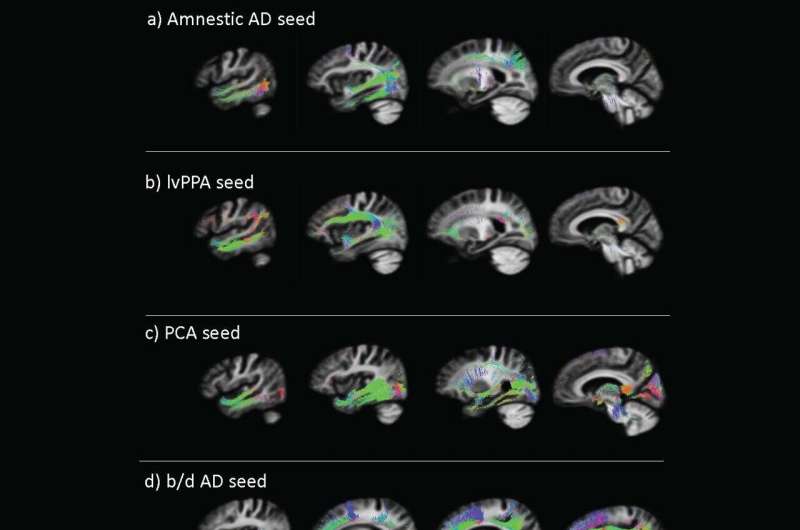
In the research, published in Science Translational Medicine, Therriault and his McGill colleagues defined the phenotypes of Alzheimer’s as amnestic, referring to memory loss; visuospatial, the inability to process information about 3D shapes; language-oriented concerns, referring to word-usage problems. This would include difficulty choosing or recalling the right word. Language problems can progress, experts say, causing impairment in patients’ ability to express themselves. A specific form of language-usage problem in Alzheimer’s is referred to as logopenic variant primary progressive aphasia, or lvPPA.
Patients with lvPPA have increasing trouble thinking of the words they want to say. As Alzheimer’s progresses, these patients have more trouble getting the words out, which eventually causes them to speak slower.
Another category outlined in the research is the behavioral and dysexecutive phenotype. Behavioral concerns can include any number of issues, covering a wide range.
The National Institute on Aging, a division of the National Institutes of Health in the United States, defines common behaviors as: Getting upset, worried or angry more easily; being depressed and uninterested in activities that were once enjoyed; hiding things or thinking that other people are hiding things; imagining things that aren’t there; pacing; wandering away from home; or showing unusual sexual behavior.
The dysexecutive phenotype refers to more of a syndrome in which an individual’s ability to multitask, organize and plan is lost. The Canadian team defined still other Alzheimer’s phenotypes: people with posterior cortical atrophy and progressive aphasia.
How the tiny protein tau plays a role in such complex Alzheimer’s phenotypes is the scientific question fueling the years-long McGill investigations. The researchers are illuminating tau’s role in the slow unraveling of the human mind.

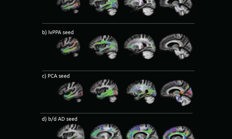
Therriault and colleagues used MRI scans to track the spread of tau in the brains of patients with four types of Alzheimer’s disease: amnestic (28 patients); behavioral/dysexecutive (15 patients); posterior cortical atrophy (19 patients), and logopenic variant primary progressive aphasia (11 patients). As a comparison, the team also performed MRI scans on 131 age-matched controls who were cognitively unimpaired.
The scientists linked each phenotype of Alzheimer’s disease to specific patterns of tau aggregation and found that tau “seeds” often appeared in areas of the brain that act as connective hubs.
Tau is actually an acronym that stands for tubulin-associated unit. There is a family of about six of these proteins, and when healthy, all are endowed with roles of maintaining the stability of microtubules, which are tough polymers, shaped like tubes. Microtubules make-up the structure of healthy cells, something cell biologists refer to as the cytoskeleton. Tau is abundant in neurons, and the brain’s cerebral cortex has the highest concentration.
But Therriault and colleagues tapped into the darker side to tau—toxic tau—aberrant proteins that are present in various neuro-inflammatory brain disorders, including Alzheimer’s, other forms of dementia and Parkinson’s disease.
“Each Alzheimer’s disease phenotype is associated with a distinct network-specific pattern of tau aggregation, where tau aggregation is concentrated in brain network hubs. In all Alzheimer’s disease phenotypes, regional tau load could be predicted by connectivity patterns of the human brain,” Therriault added. “Furthermore, regions with greater connectivity displayed similar rates of longitudinal tau accumulation.”
Tau isn’t the only deleterious protein in Alzheimer’s disease. Many people are familiar with the term “plaques and tangles” as a description of the pathology defining the Alzheimer’s brain. Plaques refer to the gummy wads of amyloid protein; the neurofibrillary tangles are the aggregates of hyperphosphorylated tau.
Although a fiery debate has raged for decades as to whether it is the plaques of amyloid-beta or the tangles of tau that trigger Alzheimer’s, neither has been declared the primary suspect. The McGill research tips the balance in favor tau, a misfolded protein with prion-like behavior.
Prions are misfolded proteins that cause a wide range of devastating mammalian brain diseases. Nobel Prize-winning neurologist Stanley Prusiner earlier this month predicted Alzheimer’s eventually would be defined as a prion-triggered disease. Prusiner, director of the Institute for Neurodegenerative Diseases and professor of neurology at the University of California, San Francisco, made the claim during the 5th Annual Constellation Forum, a symposium sponsored by Northwell Health, New York State’s largest health care system.
Previous preclinical research has suggested that tau can spread from neuron to neuron along synapses in the brain, similar to how prions spread through the brain in infected mammals, such as cattle overcome by so-called mad cow disease.
Source: Read Full Article
