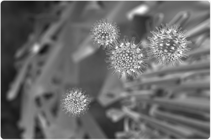
Serial block-face scanning electron microscopy (SBEM) was originally developed to analyze connectivity of axons in the brain, but has found wide applicability for many types of biological samples.
 Credit: PLRANG ART/Shutterstock.com
Conventional microscopy cannot resolve some cellular structures, and does not capture three-dimensional features of the sample. The connectivity of local networks of neurons is an example of information that could be lost in a biological tissue under an electron microscope or a light microscope.
SBEM is a type of scanning electron microscope that has an ultramicrotome mounted inside its vacuum chamber. The sample is fixed and stained and the surface is imaged by detection of back-scattered electrons. Then a thin section of about 30 nm is cut from the face of the block and it is imaged again.
Resolution of SBEM imaging
Resolution of the image depends on the lateral spatial resolution, probe depth, and slice thickness. The spatial resolution of the back-scattered electron image is determined by the probe size of the electron beam and the volume of the back scattered electron output. Resolution on the Z axis, or directly into the sample, is determined by the beam penetration depth rather than slice thickness.
SBEM is now being applied to a variety of biological samples. Kremer et al. reported being able to image features such as Arabidopsis plasmodesmata which appeared only sparsely with 2D tunneling electron microscopy.
The potential of 3D SBEM imaging not only includes visualization of complex structures, but other advantages, such as the ability to zoom from a millimeter to nanometer range and back again. This offers the possibility of capturing rare events more easily located on a larger overview, and then zooming in on those events to visualize the nanostructure.
Quantitative studies can also be carried out without the restrictions of small sample size that go with 2D electron microscopy. Functional information from a live cell connected with tissue imaging and fine-grained subcellular imaging could potentially enable the creation of 2D atlases of whole tissues or organisms.
Big data
SBEM produces an extremely large volume of data, even with a very small sample size. The computational power required to visualize and reconstruct the images is considerable. It can take several hours to reconstruct even a small data set, such as an organelle.
However, the data generated by a single experiment is highly recyclable. SBEM reveals full structural details of the cell, so data used in one investigation can be reused to answer a different question. Thus, the potential exists to create libraries of SBEM data sets for sharing and reuse by the scientific community.
Challenges
Advances in computational architecture are needed to realize the full potential of SBEM. That includes advances in reconstructing high-quality images, automated segmentation of substructures, correlation of SBEM data with light microscope data, and systems for archiving and data mining. As well, improvements are needed in the workflow of the device to integrate it with a typical life science laboratory.
Sources:
- Serial Block-Face Scanning Electron Microscopy to Reconstruct Three-Dimensional Tissue Nanostructure
- Enhancing serial block-face scanning electron microscopy to enable high resolution 3-D nanohistology of cells and tissues
- Developing 3D SEM in a broad biological context
- Serial block-face scanning electron microscopy to reconstruct three-dimensional tissue nanostructure
Further Reading
- All Electron Microscopy Content
- Electron Microscopy: An Overview
- Applications of Electron Microscopy
- Advantages and Disadvantages of Electron Microscopy
- What is Transmission Electron Microscopy?
Last Updated: Aug 23, 2018

Written by
Dr. Catherine Shaffer
Catherine Shaffer is a freelance science and health writer from Michigan. She has written for a wide variety of trade and consumer publications on life sciences topics, particularly in the area of drug discovery and development. She holds a Ph.D. in Biological Chemistry and began her career as a laboratory researcher before transitioning to science writing. She also writes and publishes fiction, and in her free time enjoys yoga, biking, and taking care of her pets.
Source: Read Full Article
